Why are the magnets used in MRI scanners supercooled?
I see many are confused about MRI magnets. Some think they're just regular magnets. MRI magnets are special. Let's learn more.
MRI magnets are supercooled to reduce electrical resistance. This allows for a stronger and more stable magnetic field[1], which is crucial for high-quality imaging in MRI scanners[2].
Keep reading to really understand the amazing world of supercooled MRI magnets.
Table of Contents
How do MRI magnets work?
I used to wonder how these magnets functioned. I didn't know their basic operation. Awareness? MRI magnets create a strong magnetic field. Let's explore.
MRI magnets work by generating a powerful magnetic field. This field aligns the hydrogen atoms in the body. Radio waves then disrupt this alignment, and the atoms' return to alignment is detected to create an image.
MRI magnets play a fundamental role in the entire MRI imaging process. At their core, they are designed to produce an extremely uniform and intense magnetic field. This magnetic field is what sets the stage for the rest of the MRI process to occur.
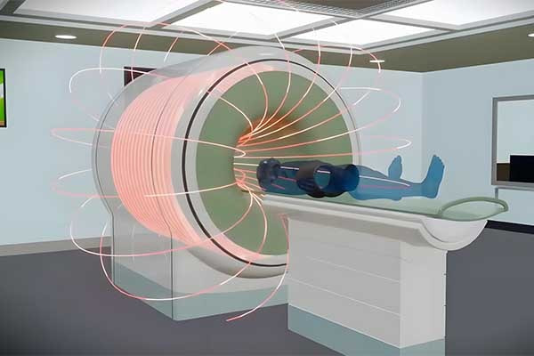
The first key aspect of how MRI magnets work is the alignment of hydrogen atoms[3]. Our bodies are mostly made of water, and water molecules contain hydrogen atoms. When a person is placed inside the MRI scanner which is essentially a large tube surrounded by the MRI magnet,, the strong magnetic field forces the hydrogen atoms' nuclei (which act like tiny magnets themselves) to align either parallel or anti-parallel to the main magnetic field.
The next step involves the use of radio-frequency (RF) pulses[4]. These pulses are sent into the body at a specific frequency. This frequency is carefully tuned to match the natural resonance frequency of the hydrogen atoms in the magnetic field, a phenomenon known as the Larmor frequency[5]. When the RF pulses are applied, they cause the hydrogen atoms to absorb energy and change their alignment. In other words, the RF pulses knock the hydrogen atoms out of their initial alignment with the main magnetic field.
Once the RF pulses are turned off, the hydrogen atoms gradually return to their original alignment with the main magnetic field. As they do this, they release the energy they absorbed from the RF pulses in the form of radio waves. These emitted radio waves are detected by the MRI scanner's receivers. The strength and timing of these detected signals vary depending on the type of tissue and the location of the hydrogen atoms within the body.
The MRI scanner then uses complex mathematical algorithms to process these detected signals and create detailed images of the internal structures of the body. Different tissues in the body, such as bone, muscle, and fat, have different concentrations of hydrogen atoms and different relaxation times (the time it takes for the hydrogen atoms to return to their original alignment). This difference in relaxation times is what allows the MRI scanner to distinguish between different tissues and create the detailed images that are so useful in medical diagnosis.
To better understand the different components[6] involved in the MRI magnet operation, we can look at the following table:
| Component | Function |
|---|---|
| Main Magnet | Generates the strong, uniform magnetic field for aligning hydrogen atoms |
| RF Coils[7] | Transmit RF pulses to disrupt hydrogen atom alignment and receive the emitted radio waves |
| Gradient Coils[8] | Create small magnetic field gradients to help localize the signals for imaging |
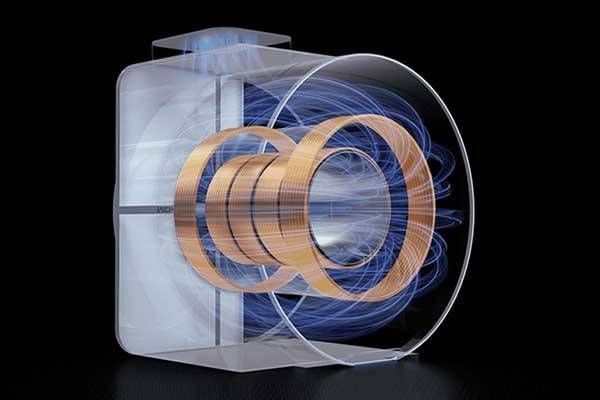
The main magnet is the most prominent part of the MRI system. It is often a superconducting magnet[9], which means it can generate extremely strong magnetic fields with very little energy consumption. Superconducting magnets are made of special materials that have zero electrical resistance when cooled to very low temperatures. This is where the supercooling of MRI magnets comes into play.
Supercooling the MRI magnets to extremely low temperatures, typically close to absolute zero (-273.15°C or 0 Kelvin), allows the superconducting materials to maintain their zero-resistance state. This is crucial because it enables the flow of large electric currents through the magnet coils. These large currents are what generate the strong magnetic fields required for MRI imaging. Without supercooling, the resistance in the magnet coils would cause energy losses in the form of heat, and it would be extremely difficult and energy-intensive to generate the required magnetic fields.
Moreover, the stability of the magnetic field is also enhanced by supercooling. A stable magnetic field is essential for accurate and consistent MRI imaging. Any fluctuations in the magnetic field could lead to image artifacts and inaccurate diagnoses. By keeping the magnet in a supercooled state, the superconducting properties remain stable, ensuring a consistent and reliable magnetic field for high-quality MRI imaging.
In addition, the type of superconducting material used in MRI magnets also affects their performance. Common superconducting materials include niobium-titanium[10] (Nb-Ti) and niobium-tin[11] (Nb₃Sn). Nb-Ti is more commonly used in clinical MRI scanners because it has a relatively high critical temperature (the temperature below which the material becomes superconducting) and is easier to manufacture. Nb₃Sn, on the other hand, can withstand higher magnetic fields but has a lower critical temperature and is more difficult to process.
How cold are MRI magnets?

Ever wondered why MRI magnets need to be supercooled? The answer lies in the physics of superconductivity and the extreme conditions required for clear medical imaging. Let’s explore the freezing temperatures that power these medical marvels.
MRI magnets are supercooled to temperatures near absolute zero (-269°C or -452°F). This extreme cold allows the niobium-titanium alloy within the magnets to become superconducting, enabling the generation of powerful magnetic fields necessary for high-quality medical imaging.
The requirement for supercooling MRI magnets stems from the principles of superconductivity[12]. When certain materials like niobium-titanium alloy are cooled to near absolute zero, their electrical resistance drops to zero. This state, known as superconductivity, allows for the creation of incredibly strong magnetic fields with minimal energy loss.
Superconducting magnets are the backbone of modern MRI technology. Traditional magnets with electrical resistance would generate heat and require massive amounts of energy to maintain the magnetic fields needed for MRI scans. Supercooling eliminates this resistance, making the process efficient and feasible for medical use.
The cooling process for MRI magnets involves liquid helium[13]. This supercooled liquid bath surrounds the magnet coils, maintaining the niobium-titanium alloy at the necessary temperature for superconductivity. The use of liquid helium is both effective and necessary, though it presents challenges such as high costs and the need for regular replenishment.
The Physics Behind Superconductivity
Superconductivity occurs when materials reach their critical temperature (Tc), below which electrical resistance vanishes. For niobium-titanium alloy, this Tc is approximately -269°C. Below this temperature, electrons form Cooper pairs, which move through the material without resistance.
This phenomenon allows MRI magnets to generate magnetic fields up to 3 Tesla, which is about 60,000 times stronger than the Earth’s magnetic field. The stronger the magnetic field, the clearer and more detailed the medical images produced.
Here’s a comparison of MRI magnet temperatures and their effects:
| Magnet Type | Operating Temperature | Magnetic Field Strength |
|---|---|---|
| Superconducting (MRI) | -269°C (-452°F) | Up to 3 Tesla[14] |
| Permanent (Neodymium) | Room Temperature | Up to 1.5 Tesla |
| Resistive | Room Temperature | Up to 0.5 Tesla |
The quest for higher magnetic fields drives ongoing research into new superconducting materials with higher critical temperatures. High-temperature superconductors[15] (HTS), which operate at -196°C (the boiling point of liquid nitrogen), offer a more cost-effective cooling solution. However, they are not yet widely used in MRI due to manufacturing challenges.
For medical imaging, the benefits of supercooled magnets are clear. Stronger magnetic fields improve image resolution, allowing doctors to detect smaller anomalies and make more accurate diagnoses. This precision is crucial in fields like neurology and oncology, where early detection can significantly impact patient outcomes.
What are the three types of MRI magnets?
Did you know there are three main types of MRI magnets? Each has unique advantages and applications in medical imaging. Let’s break down how they differ and why superconducting magnets dominate modern MRI technology.
The three types[16] of MRI magnets are superconducting, permanent, and resistive. Superconducting magnets, cooled by liquid helium, generate the strongest fields (up to 3 Tesla) and are used in most modern MRI scanners. Permanent magnets, often made of neodymium, offer lower fields but require no power. Resistive magnets are the weakest and least common, primarily used in older or low-field MRI systems.
Understanding the three types of MRI magnets requires examining their construction, cooling requirements, and applications. Each type addresses different needs within the medical imaging community, from high-resolution scans to cost-effective solutions.
Superconducting Magnets
Superconducting magnets[17] are the gold standard for MRI. They use coils of niobium-titanium alloy cooled by liquid helium[18] to near absolute zero. This allows the coils to conduct electricity without resistance, creating magnetic fields up to 3 Tesla. These magnets are expensive to install and maintain due to the need for constant helium replenishment. However, they offer unparalleled image quality and are essential for high-field MRI systems.
Permanent Magnets
Permanent magnets, often made of neodymium, require no external power or cooling. They are lighter and more compact than superconducting magnets, making them suitable for open MRI systems and locations with limited space. However, their magnetic field strength is typically lower (up to 1.5 Tesla), which can limit image resolution[19]. Despite this, they remain popular for specific applications where accessibility and cost are priorities.
Resistive Magnets
Resistive magnets[20] are the simplest and least expensive option. They use copper wire coils to generate magnetic fields but suffer from significant energy consumption and heat generation. Their field strength is typically below 0.5 Tesla, which restricts their use to low-resolution imaging. These magnets are now largely obsolete in modern MRI but may still be found in older systems or specialized research settings.
Choosing the Right MRI Magnet
The choice of magnet type depends on several factors:
Image Quality: Superconducting magnets offer the highest resolution, critical for detailed diagnostic imaging.
Cost and Maintenance: Permanent magnets require less maintenance and are more affordable, ideal for budget-conscious facilities.
Space Requirements: Permanent and resistive magnets are more compact, suitable for facilities with limited space.
Patient Comfort: Open MRI systems using permanent magnets can reduce claustrophobia for patients.
Here’s a summary of each magnet type’s suitability:
| Magnet Type | Best For | Drawbacks |
|---|---|---|
| Superconducting | High-resolution imaging | High cost, maintenance |
| Permanent | Cost-effective, open MRI | Lower field strength |
| Resistive | Low-cost, basic imaging | High energy use, weak field |
Advancements in magnet technology continue to shape the future of MRI. High-temperature superconductors and improved permanent magnet designs are pushing the boundaries of what’s possible, promising more accessible and higher-quality imaging solutions.
The choice of MRI magnet type directly impacts patient care, research capabilities, and operational efficiency. By understanding the strengths and limitations of each type, medical facilities can make informed decisions that best serve their patients and practitioners.
What is the function of the magnet in the MRI machine?
Pain in diagnosis can feel overwhelming. You want accurate results, fast. But how does a machine see what’s inside your body without cutting you open?
The magnet in an MRI machine creates a powerful magnetic field that aligns hydrogen protons in the body. These aligned protons are then used to produce detailed internal images.
When I think about MRI technology, the magnet is the first part I consider. It’s the core of the entire imaging system. The MRI magnet, typically a superconducting magnet, generates a very strong and stable magnetic field — often 1.5 to 3 Tesla. This field is what allows the scanner to align the protons in the human body, especially hydrogen, which is abundant in water and fat. Once aligned, radio waves are used to disrupt that alignment temporarily. When the protons return to their normal state, they emit signals. The MRI scanner detects these signals and turns them into images.
Main Components of MRI Magnet Function
| Component | Role |
|---|---|
| Magnet | Creates magnetic field |
| Gradient Coils | Encode spatial data |
| RF Coils[21] | Transmit and receive signals |
This system is not just powerful; it’s precise. And to maintain this precision, the MRI magnet is supercooled using liquid helium to enable superconductivity. This way, the magnet conducts electricity with zero resistance, producing the stable, strong field needed for accurate images. That’s why, as an M-Magnet manufacturer, I understand the critical role of magnetic strength and stability in medical imaging. Our advanced neodymium and MagSafe magnets may not go into MRI directly, but the principles of high performance and low loss are the same.
MRI magnets are among the strongest used in medical applications. While they are more complex than typical industrial or consumer magnets, the underlying importance of magnetic field quality is something I keep in mind every time I design a new magnet solution for a client.
Can an MRI magnet be turned off?
You might think turning off a machine is as simple as pressing a button. But is it really that simple for a powerful MRI scanner?
MRI magnets cannot be turned off like a regular device. Superconducting MRI magnets are always on unless they are deliberately quenched or shut down, which is rare and complex.
In most MRI machines, the magnet is designed to be always active. This is especially true for superconducting magnets, which are cooled with liquid helium to near absolute zero. Once current flows through the coil, it circulates without resistance and creates a persistent magnetic field. This type of setup means there is no power supply constantly feeding the magnet. Instead, the magnetic field stays active until something causes it to stop—either a quench or a controlled shutdown. Quenching is when the magnet suddenly loses its superconducting state and rapidly warms up, releasing helium gas and collapsing the field. It’s expensive, risky, and only done in emergencies.
Methods of Deactivating MRI Magnets
| Method | When Used | Risks |
|---|---|---|
| Emergency Quench[22] | Accidents or fire | Helium loss, equipment damage |
| Controlled Ramp Down | Maintenance or decommissioning | Takes days, costly |
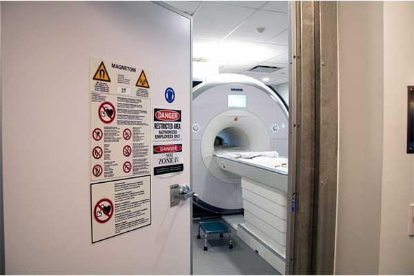
I’ve often been asked if a strong magnet like an MRI magnet can be turned off when not in use. From an engineering point of view, the answer is no—at least, not easily. This is one of the reasons why MRI environments are always treated with extreme caution. Since the magnetic field is always present, any ferromagnetic object can become a projectile if brought too close.
At M-Magnet, we handle high-performance magnet materials like neodymium that can generate very high magnetic forces. Even though they are permanent magnets and not superconducting like in MRI, safety and magnetic stability[23] are still a top concern. Designing custom solutions for clients in America and Europe has shown me how important magnetic behavior is in various industries, including healthcare.
We don’t directly make MRI magnets, but the understanding of magnet strength, cooling, and control is still part of our design thinking. The lessons learned from MRI magnets also help us optimize our products for precision and reliability in MagSafe and wireless charging applications, where magnetic field behavior matters just as much.
Why are MRI magnets so strong?

MRI scanners need very strong magnetic fields[24]. These strong fields help create clear images of the inside of the body. The superconducting magnets inside MRI scanners are made with special wires that lose almost all electrical resistance when cooled to very low temperatures, creating a very strong and stable magnetic field without needing a constant power supply.
MRI magnets are very strong because they use superconducting wires cooled to extremely low temperatures. This superconductivity allows a large electrical current to flow with almost no resistance, generating powerful magnetic fields needed for clear MRI images[25].
The Science Behind Superconducting Strength
To understand why MRI magnets are so strong, we need to look at how they work and the role of supercooling. Regular electromagnets create a magnetic field by passing an electric current through a coil of wire. The strength of the magnetic field depends on the amount of current and the number of turns in the coil. However, the wire has electrical resistance, which causes some of the electrical energy to be lost as heat. To get a very strong magnetic field, a very large current is needed, which leads to a lot of heat and high energy consumption.
Superconducting magnets solve this problem by using materials that become superconducting at extremely low temperatures, typically close to absolute zero (-273.15 °C or 0 Kelvin). In this superconducting state, the electrical resistance of the wire drops to virtually zero. This means that once an electric current is started in a loop of superconducting wire, it can flow continuously without any energy loss. Therefore, a large current can be maintained, generating a very strong magnetic field without the need for a constant power supply to overcome resistance. This is crucial for MRI machines, which require strong and stable magnetic fields for high-quality imaging.
The materials commonly used in MRI magnets are special alloys, such as niobium-titanium (Nb-Ti)[26] and niobium-tin (Nb3Sn)[27]. These materials exhibit superconductivity at cryogenic temperatures, which are achieved by immersing the magnet coils in liquid helium. Liquid helium has a boiling point of about 4.2 Kelvin (-268.95 °C), making it ideal for cooling superconducting magnets.
The strong magnetic fields produced by these superconducting magnets interact with the atomic nuclei in the patient's body. Specifically, they align the protons within the nuclei. Radio waves are then emitted into the body, which temporarily knock these aligned protons out of their equilibrium state. As the protons return to their original alignment, they emit signals that are detected by the MRI scanner. These signals are then processed by a computer to create detailed images of the body's internal structures. The stronger the magnetic field, the more aligned the protons become, and the stronger the emitted signals, resulting in clearer and more detailed images.
The stability of the magnetic field is also very important for MRI imaging. Superconducting magnets can maintain a very stable magnetic field over long periods because the current flows continuously without resistance. This stability ensures that the images produced are consistent and free from artifacts that could be caused by fluctuations in the magnetic field.
As a neodymium magnet manufacturer and a MagSafe magnet factory, M-Magnet Company understands the importance of strong and stable magnetic fields in various applications, although the specific technology used in MRI magnets differs from our standard product lines. The principles of generating strong magnetic fields efficiently are fundamental in both areas.
| Property | Regular Electromagnet | Superconducting Magnet |
|---|---|---|
| Operating Temperature | Room temperature or slightly elevated | Cryogenic temperatures (near absolute zero) |
| Electrical Resistance | Significant | Virtually zero[28] |
| Current Requirement | High current needed for strong field | High current can be sustained with no loss |
| Energy Consumption | High, due to resistive losses | Very low, once the current is established |
| Magnetic Field Strength | Limited by heat generation and power supply | Very high and stable |
| Cooling System | Typically air or water cooling | Liquid helium cooling |
| Cost of Operation | High, due to electricity consumption | Relatively low, after initial cool-down |
The use of superconducting magnets is a key technology that enables MRI scanners to produce high-resolution images of the human body. The ability to generate and maintain very strong magnetic fields efficiently is essential for this medical imaging technique.
Conclusion
In conclusion, MRI magnets are supercooled to enable the creation of a strong and stable magnetic field. They work by aligning hydrogen atoms in the body using a powerful magnetic field and then using RF pulses to create images. Supercooling is vital for reducing resistance and ensuring the magnet functions effectively. Understanding these aspects helps us appreciate the technology behind MRI scanners, which are so important in modern medical diagnosis.
Note:
[7]Offers a practical guide to RF coils in MRI, detailing their types and functions.↪
[8]Explains the function and design of gradient coils in MRI, essential for spatial encoding.↪
[25]Explain how powerful magnetic field is required for a high resolution images on MRI scanners.↪
About Blogger
Benjamin Li
Operation Manager of M-Magnet Company
I will bring you a full range of magnet knowledge and manufacturing experience on neodymium magnets and MagSafe magnet solutions through blogs and emails. I'm not an expert yet in magnets, but we have a whole team to help you solve technical issues, design drawing details, compatibility suggestions from magnetic assemblies, magnet purchasing and many other customized magnet solutions from China. You can follow my blogs on knowledge sharing or contact me for your own magnet solutions. We will always do the best.